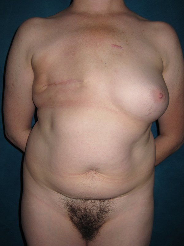
Before and after composite DIEP flap and breast implant reconstruction in a 46-year-old breast cancer survivor. She had a prior right mastectomy, chemotherapy and radiation therapy. She wanted to use her abdominal tissue for reconstruction, and to regain her prior breast fullness that she had lost after breastfeeding her children.
She had enough abdominal skin and fat to create a large right breast reconstruction using a DIEP flap. Tissue was microsurgically transplanted from her lower abdomen to her right chest, disconnecting and then reconnecting one tiny artery and two tiny veins (2-3 millimeters) under the microscope.
A smooth, round saline-filled permanent but postoperatively adjustable breast implant was placed under the left breast in the subglandular position, on top of the muscle. After surgery, the patient gained a little weight, which increased the volume of her DIEP flap – the fat cells do not realize they are now located in the breast, not the tummy! The left implant volume was increased by adding additional saline to the implant to achieve the best possible symmetry.
At a secondary procedure, right nipple reconstruction was performed using the nipple-sharing technique, where a portion of the left nipple was transplanted to the right breast reconstruction as a free nipple graft. Medical tattoo created a new areolar circle. The left implant port was removed at that time as well, and the saline implant remained in place.
Follow up photos are shown 4 years after surgery. Thanks to microsurgery and her breast reconstruction, she no longer thinks daily about her breast cancer and the associated loss. She has resumed a normal active lifestyle (sans breast prosthesis) with a positive body image.


*All photos are actual patient photographs and are for illustrative purposes only. Individual results may vary.