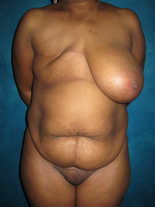
Before and after right delayed breast reconstruction with the DIEP flap in a 43-year-old breast cancer survivor. She was treated with a right non-skin-sparing mastectomy, chemotherapy and right chest wall radiation. She was not offered reconstruction, and she took her time researching her options before choosing a DIEP flap and for our team to care for her.
Her ideal reconstruction technique involved autogenous tissue from her own body to help bring new healthy tissue to the radiated field. The option of a tissue expander and implant had a high risk of complications including infection, capsular contracture, skin loss and potential loss of the implant because of past significant radiation that she was not willing to accept.
For the DIEP flap, skin and fat was microvascularly transplanted from her lower abdomen to her right chest. The procedure takes anywhere from four hours for one breast to 8 hours for both breasts to perform, using a two-co-surgeon team approach at the hospital where Reconstructive Microsurgery was developed, and where Dr. Horton completed her final year of Fellowship training in Microsurgery in 2006.
An immediate right nipple-areola reconstruction was performed on the left side using the “nipple-sharing” technique, where a piece of the left nipple was transplanted to the right as a free nipple graft. In addition, some of the left areola skin was transplanted as a full-thickness skin graft directly to the right breast reconstruction.
A balancing breast reduction and lift was performed on the left side. She did not want her breasts to be too small, so the left breast was created the estimated size that she preferred, and the right breast was reconstructed as large as possible, given her radiated chest and vascular details of the DIEP flap.
Follow up photos are shown 8 months after surgery. Once she heals further, she will be a candidate for an implant beneath her right DIEP flap to augment her reconstruction further and achieve greater symmetry and/or free fat grafting to add additional volume to her flap.


*All photos are actual patient photographs and are for illustrative purposes only. Individual results may vary.