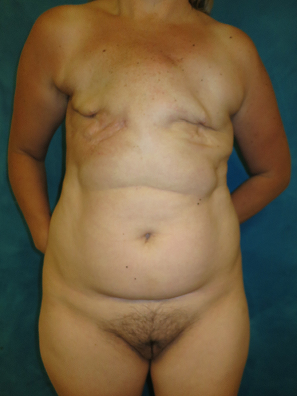
Before and after bilateral delayed DIEP flap breast reconstruction in a 45-year-old breast cancer survivor. She had submuscular tissue expanders placed at the time of her mastectomies which became infected during chemotherapy, when her immune system was compromised, requiring their removal. Two years later, when she had finished chemotherapy and radiation therapy, she was ready to revisit breast reconstruction.
Because her right chest wall tissue had been radiated, a flap (using the body’s own tissue for reconstruction) was her best option. Thankfully, she had sufficient lower abdominal skin and fat to build two new breasts. Bilateral DIEP flaps were transplanted microvascularly from her lower abdomen to her chest as “free flaps” (moving the tissue free in the air versus using a muscular pedicle as a method to preserve the blood supply).
Free flap reconstruction involves dissecting tiny (2–3-millimeter diameter) arteries and veins, dividing them at the donor site, and reconnecting them under the microscope at the recipient site. We select the internal mammary artery and vein as two connections, and a branch of the axillary vein in the armpit region as a second venous anatstomosis in all cases in order to preserve the best possible venous outflow of each flap.
One year later, nipple and areola reconstructions were performed using the local flap technique and medical tattoo at the same time as free fat grafting, used to further contour her abdomen and flanks, and add a small amount of additional volume to her flaps.
Follow up photos are shown 4 years after DIEP flap reconstruction.


*All photos are actual patient photographs and are for illustrative purposes only. Individual results may vary.