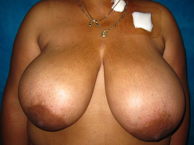
Before and after right lumpectomy with positive margins requiring a wide re-excision and local tissue rearrangement in a 41 year old woman with breast cancer. Following neoadjuvant chemotherapy, she was a candidate for a breast reduction on both sides and was grateful she could have her repeat lumpectomy and reconstruction all in one surgery. She just had her chemo port removed, as evidenced by the left chest bandages.
The Breast Surgeon performed wide excision of the lumpectomy, which had negative margins. Due to the nature of her breast cancer, it was known that she required postoperative radiation therapy. The remainder of the right breast was rearranged using the breast reduction/lift technique. A left breast reduction was performed at the same surgery, and liposuction removed excess fat from the axillary rolls (armpits and bra rolls).
It is known that radiation will cause breast shrinkage of 5% to 20%, depending on the dose of radiation and the body’s response. We describe radiation change like putting a wool sweater in the dryer on high for just a few minutes (whoops!) – The sweater will keep its shape, but the entire garment will shrink a little in all dimensions.
The same thing happens with a breast that receives radiation. The entire breast will shrink, and the nipple will rise a few centimeters. Therefore, when it is known that radiation is required after breast reconstruction, the reconstructed breast is made 5-20% bigger with a lower nipple than the non-radiated breast to allow for symmetry once radiation is complete. It can take 6 months to a year for radiation changes to become evident.
Follow up photos are shown a year and a half after surgery, and 14 months after radiation was completed. Interestingly, the radiated scars look nicer than the non-radiated scars! This is because radiation is a treatment for keloid scars – it limits collagen deposition.


*All photos are actual patient photographs and are for illustrative purposes only. Individual results may vary.