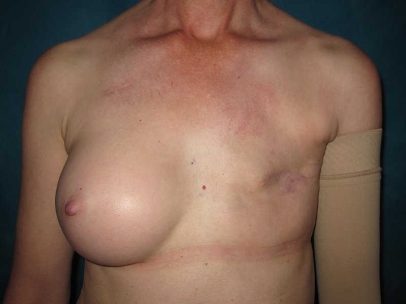
Before and after left breast reconstruction with a TUG flap and secondary sub-flap implant in a 53-year-old breast cancer survivor. Following her left mastectomy and chemotherapy, she required radiation to her chest wall.
Although her tissue expander on the left had been placed at the time of her mastectomy, it got infected after chemo and radiation and required removal. She previously had a right breast augmentation and wanted to keep this implant – she liked the fullness and roundness of her breast.
A two-stage procedure was planned for her reconstruction. Since she had very little abdominal skin and fat and she had already failed an implant-based reconstruction, she needed tissue from elsewhere on her body. She had enough upper inner thigh skin and fat to use – hence our decision to use a TUG flap.
A TUG flap was transplanted microsurgically from her right upper inner thigh to the left chest wall. The TUG flap has the advantage of enabling immediate nipple and areola reconstruction at the same time as the flap by accentuating the standing cone created when the flap is coned on the back table. More information can be found here about this technique.
Six months later, a smooth round permanent and postoperatively adjustable breast implant was placed under the flap and inflated to match the right augmented breast. Follow up photos are shown 1 year after her TUG flap and 3 weeks after implant placement. A small circle band aid is over the remote implant port – this port will stay in place up to a year to ensure the absolute best symmetry of her left breast reconstruction to her right and can be removed in the office under local anesthesia at any time.


*All photos are actual patient photographs and are for illustrative purposes only. Individual results may vary.